DNA 复制叉机制图
中文完整提示词
一张 DNA 复制叉侧视扁平矢量图。亲代双链螺旋从左侧以深蓝与青绿色进入;解旋酶为六聚体灰色环;前导链(深蓝)上钴蓝 C 形 Pol III 夹持滑动,合成青色新链;滞后链(青绿)上三段淡金色冈崎片段,每段以珊瑚色 RNA 引物起始,绿色椭圆 DNA 连接酶封口,RNase H 移除一段引物。叉前方 Topo I 以 8 字结释放超螺旋。领线细标注,右上角色码图例,白色背景,无阴影。
English full prompt
A detailed flat vector diagram of an active DNA replication fork, viewed from the side as a close-up cross-section. The parental double helix enters from the left as two intertwined strands in complementary navy and teal. At the fork junction, a helicase enzyme is shown as a hexameric ring (6 pale grey subunits) actively unwinding the strands. The leading strand (navy, top) runs continuously rightward, with DNA polymerase III depicted as a C-shaped clamp in cobalt blue sliding along it, reading 3′→5′ and synthesising 5′→3′ with a growing new strand in cyan. The lagging strand (teal, bottom) runs in the opposite direction; three Okazaki fragments in pale gold are shown, each starting with a short RNA primer in coral and being extended by a second pol III clamp. RNase H removes a primer in one fragment, and DNA ligase (a green oval) seals the nick. Topoisomerase I ahead of the fork is shown as a figure-8 knot releasing a supercoil. All components are annotated with thin leader lines and 9 pt labels. Colour-coded legend at top-right. White background, no drop shadows.
相关案例
-
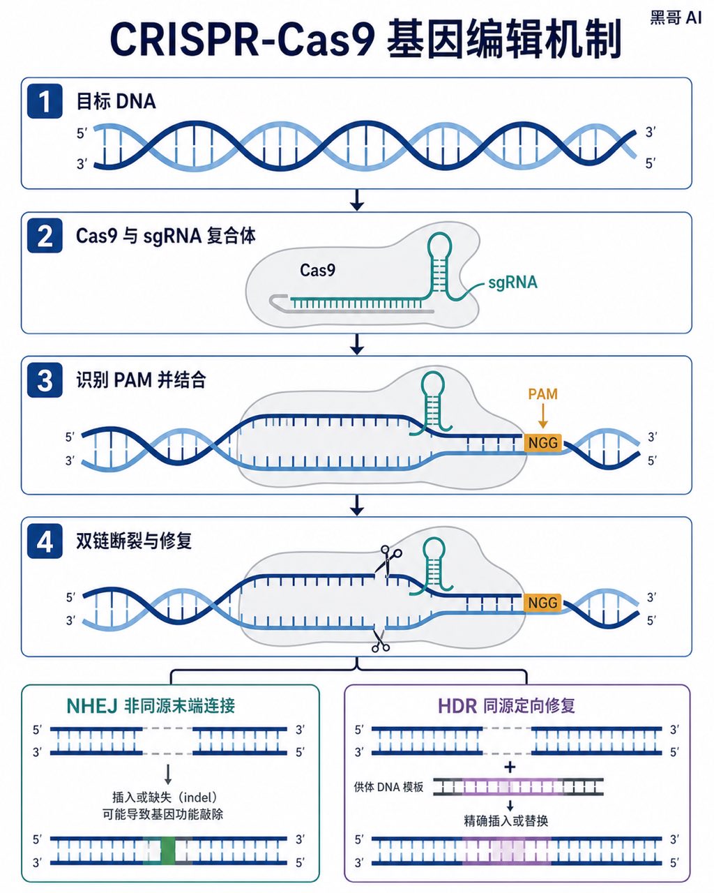
CRISPR-Cas9 基因编辑机制图
生成一张 4:5 竖版「CRISPR-Cas9 基因编辑机制图」案例图,所属分类为「科研与学术图」。四格扁平矢量图,展示 CRISPR-Cas9 切割 DNA 的全过程,深蓝螺旋、灰色 Cas9 蛋白、琥珀色 PAM 位点及红绿结果分支。画面需要完整呈现上述主体、构图、配色、材质、光线和整体风格;可见文字以自然简体中…
-
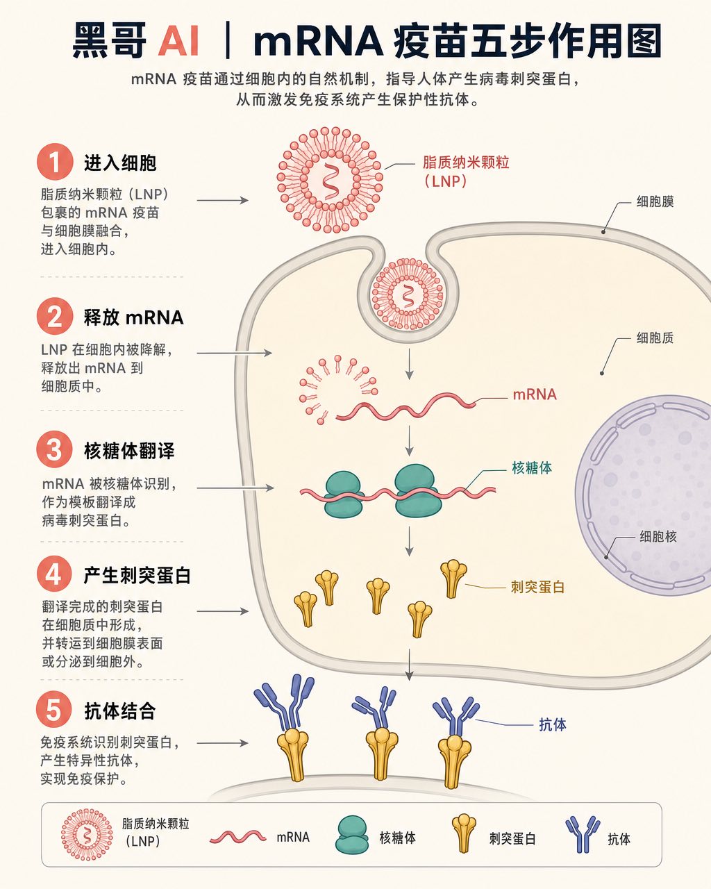
mRNA 疫苗细胞作用机制
一张教科书风格的等距信息图,展示 mRNA 疫苗在人体细胞内的分步作用。背景米白,脂质纳米颗粒(LNP)为珊瑚粉,核糖体为青绿色,刺突蛋白为金色。从 LNP 进入细胞膜,到 mRNA 释放、核糖体翻译刺突蛋白,再到靛蓝 Y 形抗体结合刺突蛋白,共五步,珊瑚色圆形数字徽章标注步骤,细箭头连接,无阴影扁平风格,适合 Sc…
-
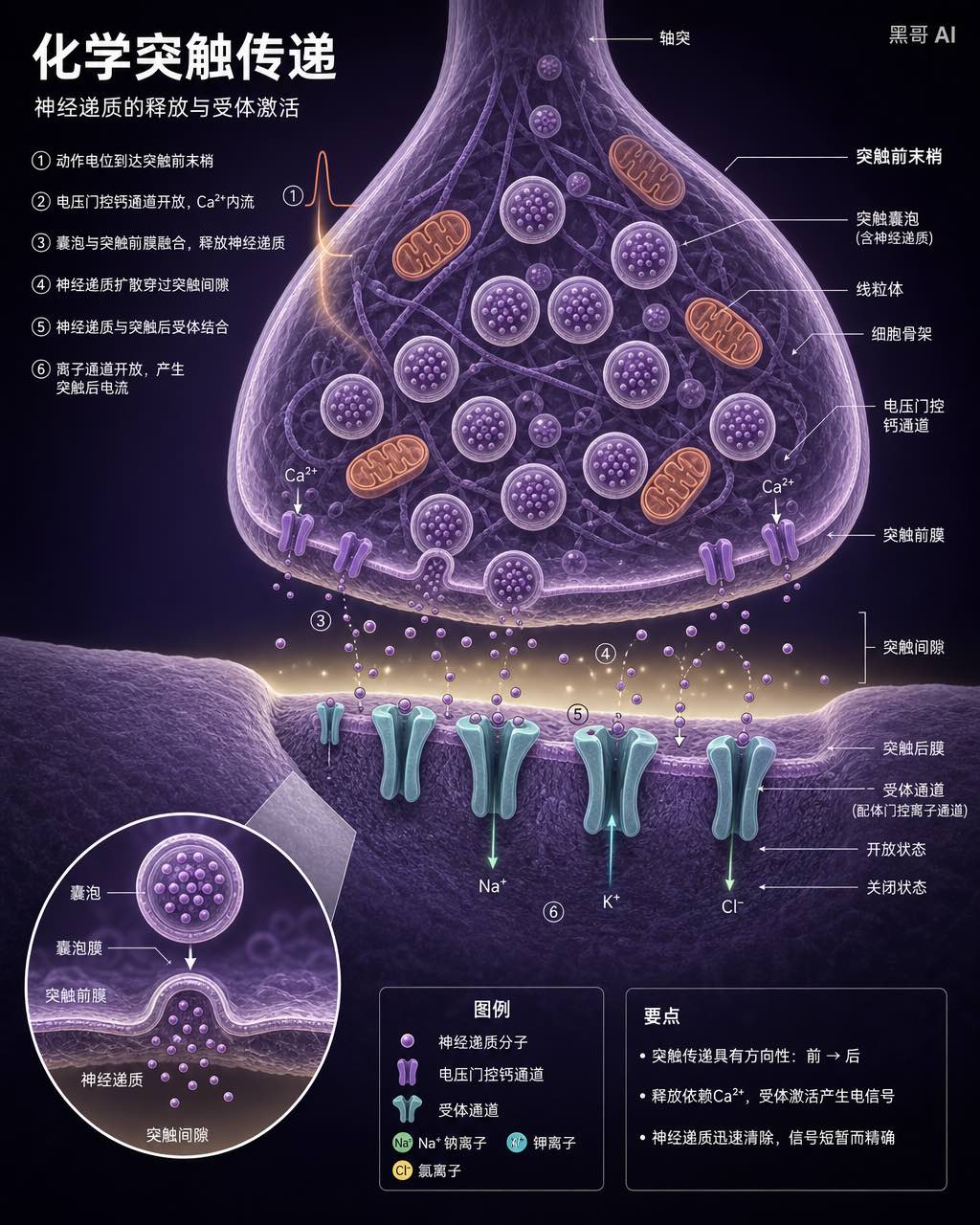
神经突触横截面图
一张高端神经科学教科书风格的化学突触横截面图。上半部为突触前末梢,布满淡紫色囊泡(内含神经递质小点)与暖橙色线粒体;突触间隙为极淡黄色细缝,神经递质分子飞渡其中;下半部突触后膜嵌有青绿色 Y 形受体通道,部分开放部分关闭,标注微小离子流箭头。小圆形插图放大一个囊泡融合事件。深紫背景,细领线标注,9pt Helveti…
-
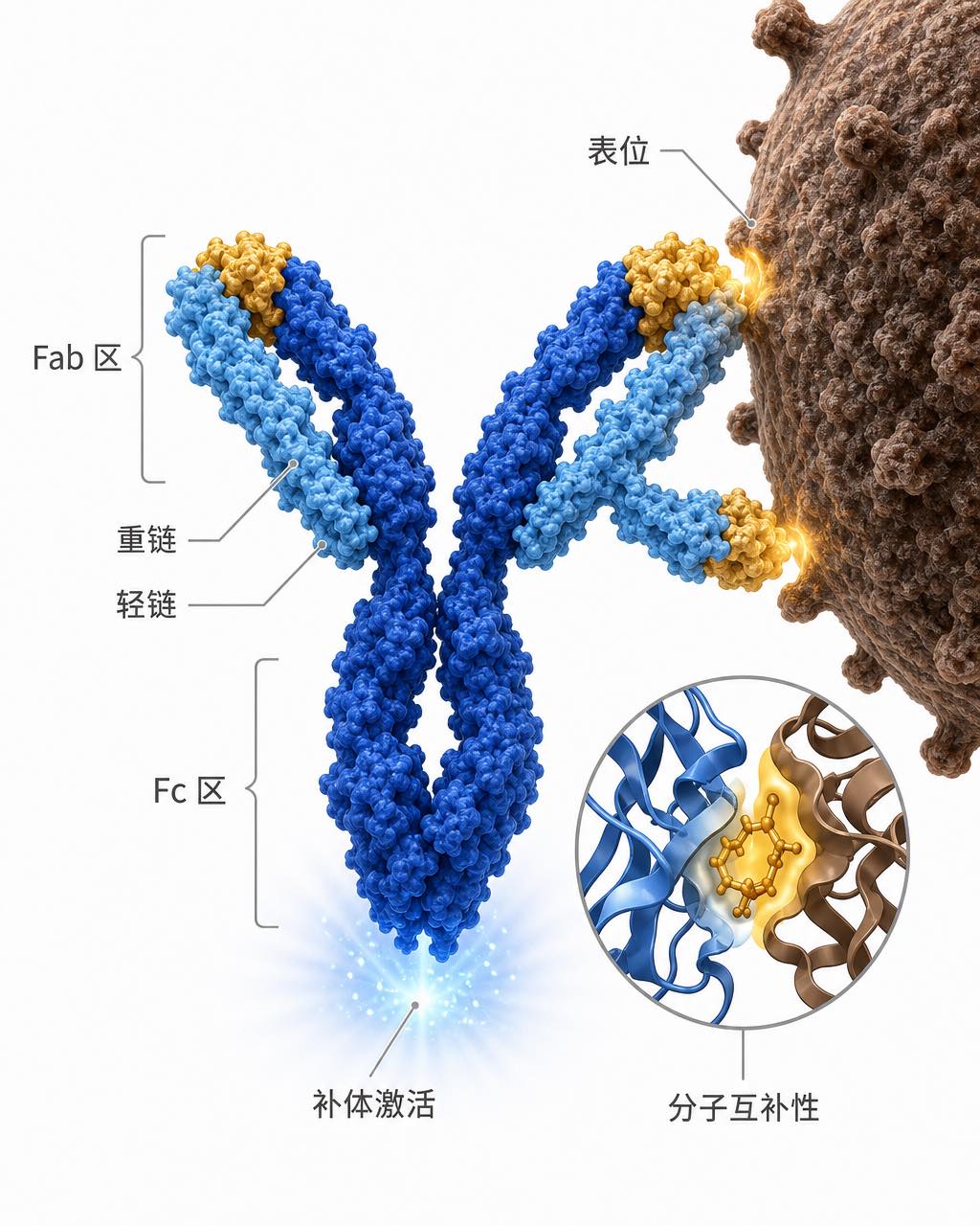
抗体-抗原结合免疫图
一张免疫学讲义风格的 IgG 抗体结合抗原示意图。深钴蓝重链与天蓝轻链构成经典 Y 形,Fab 区末端与病原体(棕褐色粗糙衣壳弧面)上的表位形成锁-钥结合。Fc 区底部微发光暗示补体激活。右下角圆形插图以 ribbon-diagram 风格展示一个结合口袋的分子互补性。白色背景,金色高亮结合区,细灰色领线标注,整洁无…