Protein Folding 3D Ribbon Diagram
English full prompt
A publication-quality 3D ribbon diagram of a globular protein showing all secondary structure elements, rendered in a PyMOL-inspired but stylistically clean aesthetic. The protein occupies the centre of a white canvas in a slightly rotated frontal view. Alpha helices are shown as wide spiral ribbons in deep cobalt blue, with each turn clearly defined. Beta sheets are depicted as flat broad arrows in warm gold pointing in the direction of the polypeptide chain, with anti-parallel sheets forming a curved beta barrel in the core. Loop regions connecting secondary structures are shown as thin silver tubes. The N-terminus is labelled "N" in green and the C-terminus "C" in red, with a faint rainbow gradient from blue (N) to red (C) along the backbone to indicate chain directionality. A disulfide bridge between two cysteine residues is shown as a short orange stick bond. The active site pocket is indicated by a dashed oval with an arrow pointing to it labelled "Active Site". Below the protein, a three-panel legend shows: α-helix, β-sheet, loop symbols. Background: white, with very subtle light grey drop shadow under the protein for depth. Scientific illustrative style with no photorealistic texturing.
中文完整提示词
一张发表质量的球蛋白 3D 带状图,PyMOL 风格但更简洁。深钴蓝螺旋宽带表示 α-螺旋,暖金宽箭头表示 β-折叠(形成核心 β-桶),银色细管表示环区。链方向以蓝(N 端绿色标 「N」)到红(C 端红色标 「C」)彩虹渐变。橙色短棒键表示二硫键。虚线椭圆加 「Active Site」 箭头标注活性口袋。底部三格图例展示 α/β/环符号。白色背景,轻微灰色投影增加深度,无写实贴图。
Related cases
-
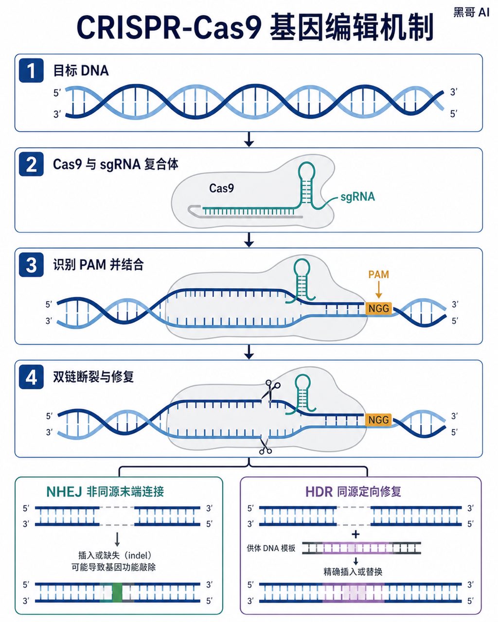
CRISPR-Cas9 Gene Editing Mechanism
A Nature-journal-quality flat vector diagram illustrating the CRISPR-Cas9 gene-editing mechanism. The composition is horizontal, spanning roughly A4 landscape, divided into four s…
-
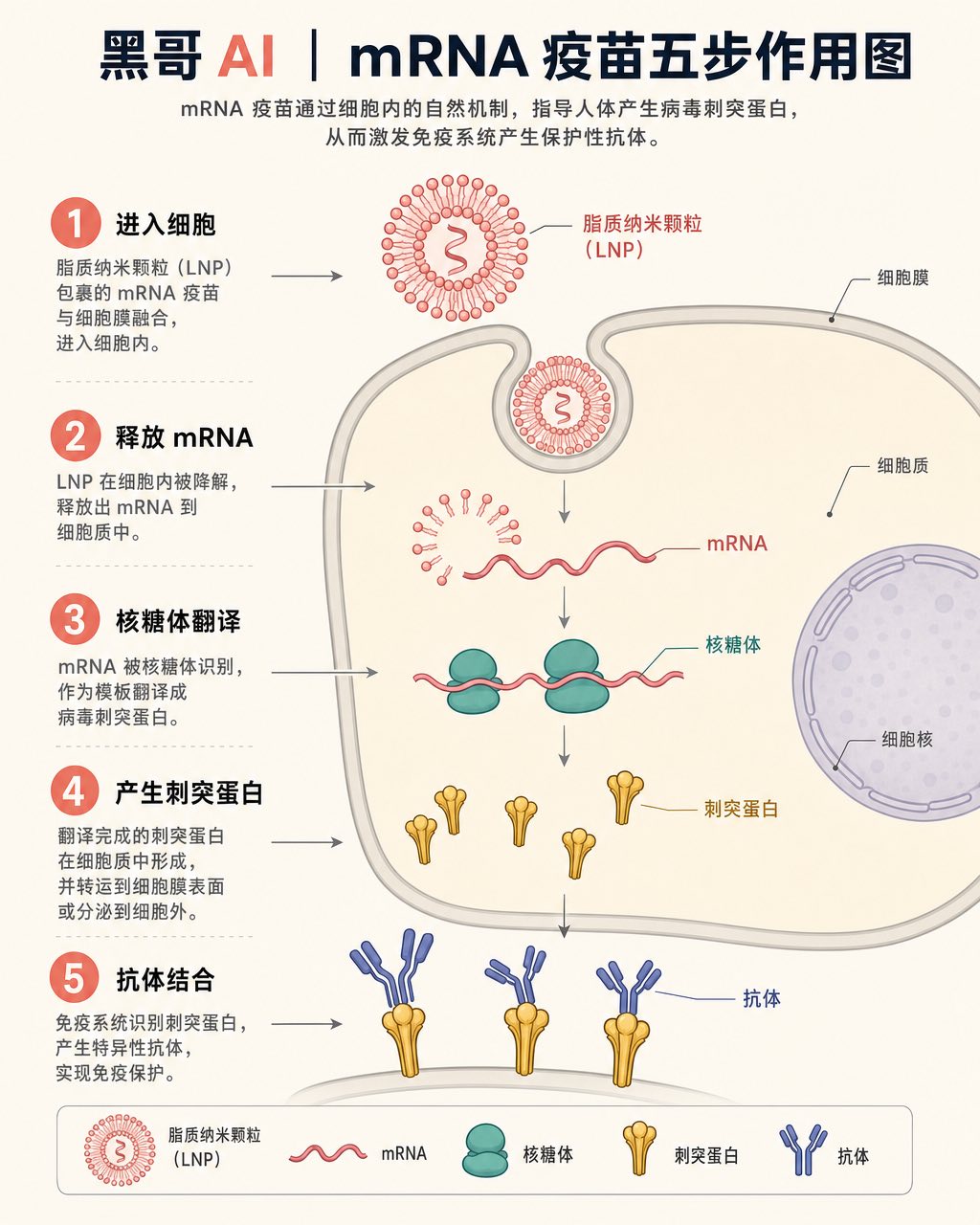
mRNA Vaccine Cellular Action
A textbook-flat isometric infographic showing the step-by-step action of an mRNA vaccine inside a human cell. The scene is rendered in a soft clinical palette: off-white backgroun…
-
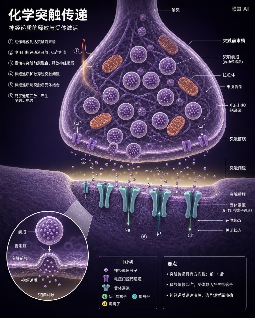
Neuron Synapse Cross-Section
A precise anatomical cross-section illustration of a chemical synapse between two neurons, rendered in the style of a high-end neuroscience textbook. Viewpoint: close-up transvers…
-
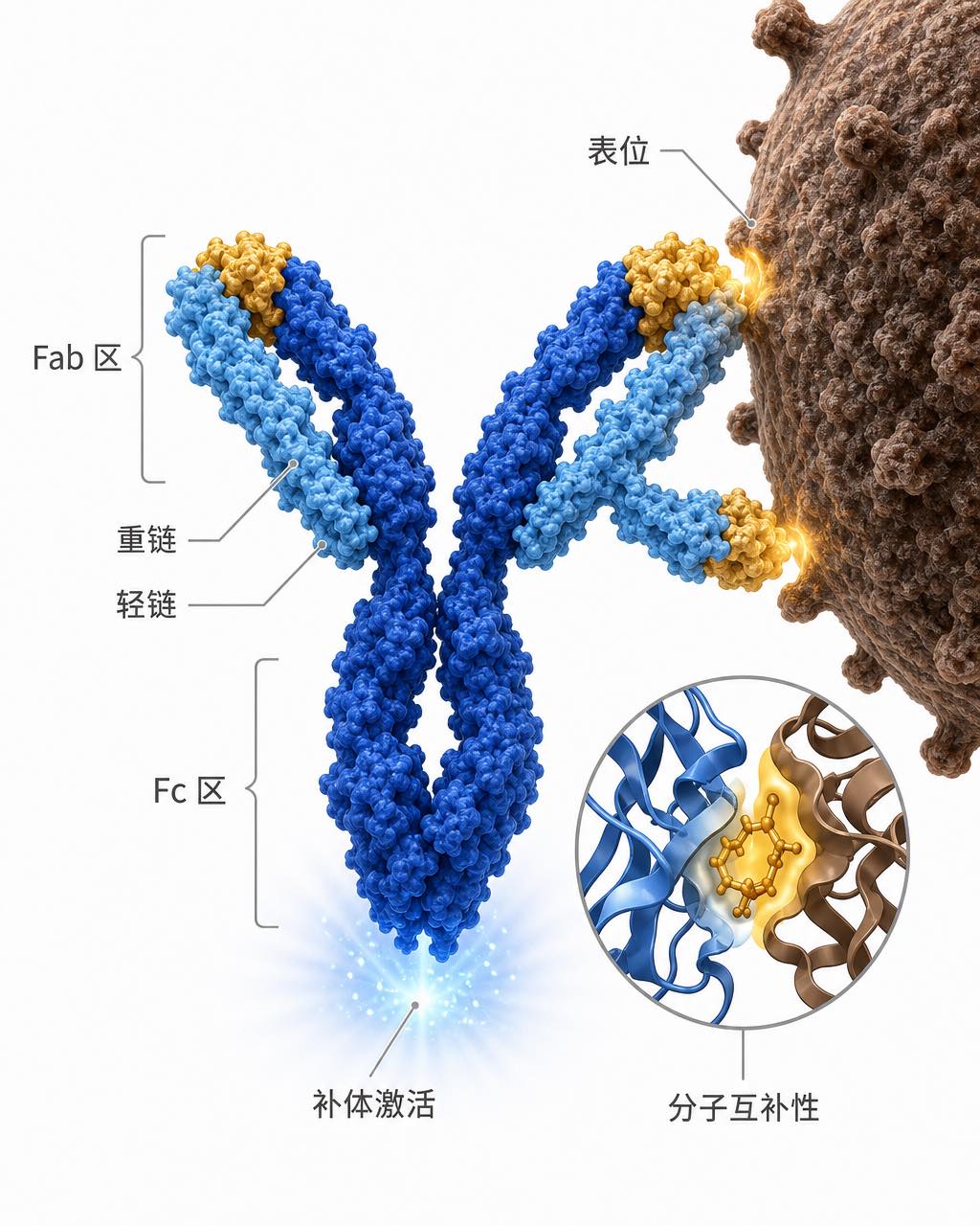
Antibody-Antigen Binding Diagram
A clean scientific illustration of an IgG antibody binding to a surface antigen on a pathogen, suitable for an immunology lecture slide. The antibody is rendered in a semi-3D ribb…