DNA Replication Fork Mechanism
This is a Scientific & Academic Figures AI image prompt case, leaning toward Flat, Line Art. Copy the full prompt below and paste it into a free GPT image generator to create your own image with GPT Image-2 — swap the subject, brand, or aspect ratio as needed.
English full prompt
A flat editorial diagram of a DNA replication fork, viewed from slightly above and to the left. The parental double helix (two intertwined blue-and-teal ribbons) unwinds at the fork point where a helicase enzyme (red ring shape) is illustrated mid-unwinding. The leading strand template (blue) is copied continuously by DNA polymerase III (dark blue teardrop), with the new strand (light cyan) growing smoothly rightward. The lagging strand template (teal) is looped back and copied in Okazaki fragments (short dashed cyan segments with gaps); primase (magenta diamond) marks the RNA primers. SSB proteins (small pale-yellow ovals) stabilize the single-stranded regions. Labels with thin hairline arrows: "Helicase", "Leading strand", "Lagging strand", "Okazaki fragment", "Primase", "SSB". Ivory background, flat colors, no gradients, 8 pt sans-serif labels.
中文完整提示词
一幅扁平编辑风格的 DNA 复制叉示意图,略从左上方俯视。亲本双链(蓝色与青色交织带)在解旋酶(红色环形)处解开。前导链模板(蓝色)由 DNA 聚合酶 III(深蓝泪滴形)连续合成新链(浅青色,向右延伸);后随链模板(青色)向后弯曲,以冈崎片段(虚线青色短段)不连续合成;引发酶(品红色菱形)标注 RNA 引物。SSB 蛋白(浅黄色小椭圆)稳定单链区域。象牙色背景,纯色无渐变,8 pt 无衬线标签。
Related cases
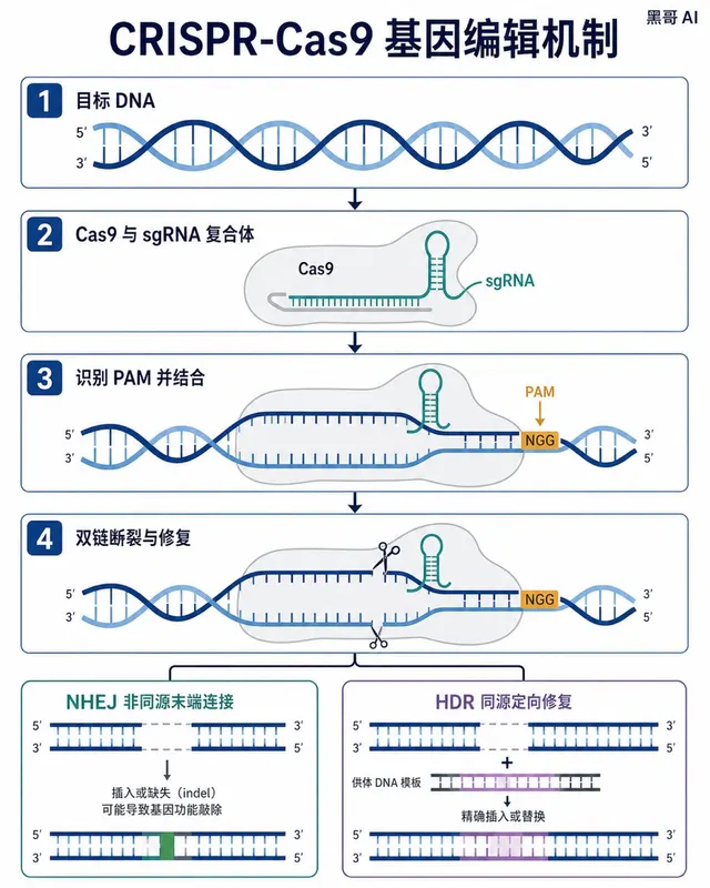
CRISPR-Cas9 Gene Editing Mechanism
A Nature-journal-quality flat vector diagram illustrating the CRISPR-Cas9 gene-editing mechanism. The composition is horizontal, spanning roughly A4 landscape, divided into four s…
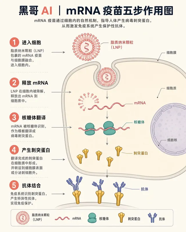
mRNA Vaccine Cellular Action
A textbook-flat isometric infographic showing the step-by-step action of an mRNA vaccine inside a human cell. The scene is rendered in a soft clinical palette: off-white backgroun…
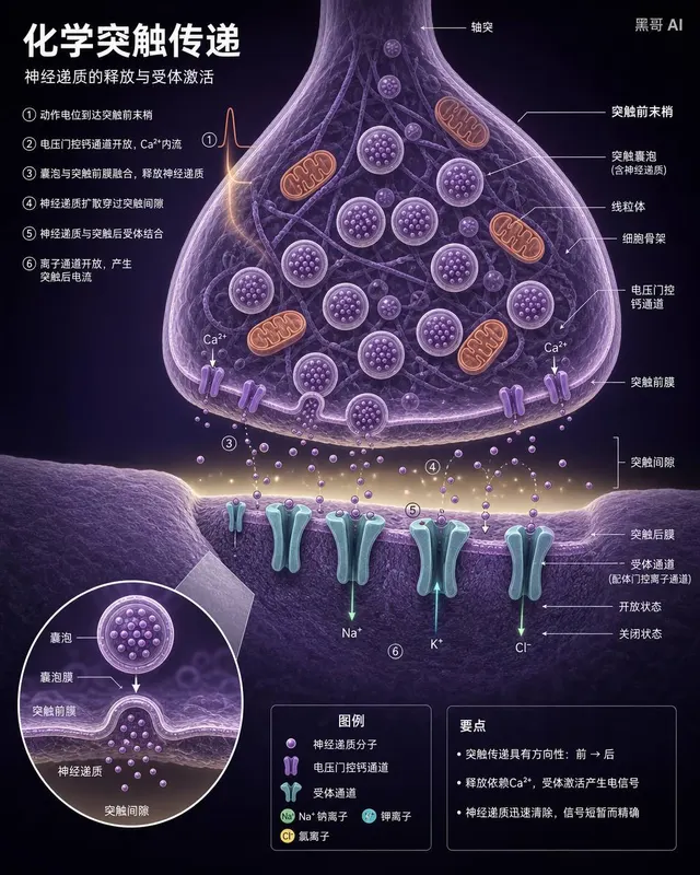
Neuron Synapse Cross-Section
A precise anatomical cross-section illustration of a chemical synapse between two neurons, rendered in the style of a high-end neuroscience textbook. Viewpoint: close-up transvers…
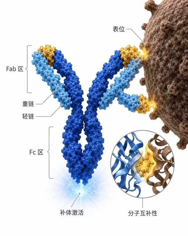
Antibody-Antigen Binding Diagram
A clean scientific illustration of an IgG antibody binding to a surface antigen on a pathogen, suitable for an immunology lecture slide. The antibody is rendered in a semi-3D ribb…